Overview
BrainNet Viewer is a brain network visualization tool for imaging connectomics, which was developed by Dr Mingrui Xia at the Imaging Connectomics Lab at Beijing Normal University.
The BrainNet Viewer can help researchers to visualize topological patterns of structural and functional brain networks derived from different imaging modalities (e.g., structural MRI, functional MRI, diffusion MRI, EEG/MEG, PET, and fNIRS) in different species (e.g., human, monkey, and rat) in a quick, easy, and flexible way. The BrainNet Viewer works well with a variety of imaging analysis tools such as Gretna and PANDA.
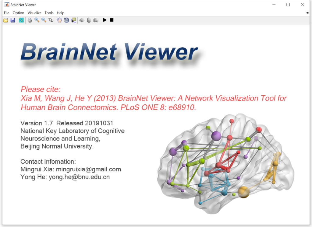 Fig. 1 A graphical user interface of BrainNet Viewer
The BrainNet Viewer includes several important features as follows:
An open-source, Matlab-based software package with a graphical user interface (GUI), illustrating connectomes as ball-and-stick models (Fig. 1);
Run on a variety of operating systems, including Windows (XP, 7, 8 and Server versions, Microsoft Corp., Redmond, WA, US), Linux (Ubuntu, CentOS, etc.) and Mac OS in both 32- and 64-bit versions;
Display brain connectomes derived from various imaging modalities (e.g., structural MRI, functional MRI, diffusion MRI, EEG/MEG, PET, and fNIRS) in different species (e.g., human, monkey, and rat) (Fig. 2);
Display brain connectomes in multi-views; display combinations of brain surface, nodes and edges; adjust properties of network elements (i.e., nodes and edges); map the volume image to brain surface; support various types of image format exporting and video making; provide interactive operations, such as zoom and rotate; and illustrate the interactions between two brain networks (Fig. 3);
Produce more than 20 cover figures in several high-profile journals such as Biol Psychiatry, Ann Neurology, J Neurosci, Cereb Cortex, Neurology, and Hum Brain Mapp (Fig. 4).
Cited by more than 1400 articles, which include many high-profile journal articles published in Nat Rev Neurosci, Lancet Neurol, Trends Cogn Sci, JAMA Oncol, Neuron, Lancet Psychiatry, Nat Commun, Trends Neurosci, Mol Psychiatry, Sci Adv, PNAS, and Brain.
 Fig. 2 Brain networks and maps derived from various imaging modalities in different species that can be generated using BrainNet Viewer
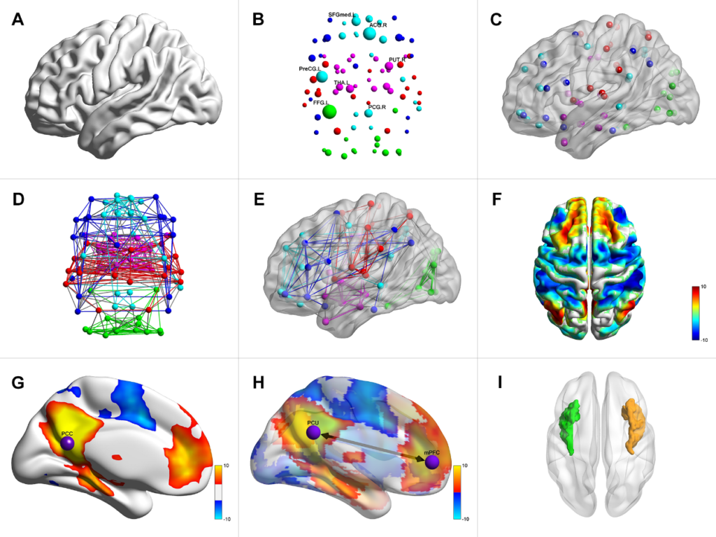 Fig. 3 Various styles of brain networks and maps that can be generated using BrainNet Viewer
 Fig. 4 Several selected cover illustrations that are generated using BrainNet Viewer
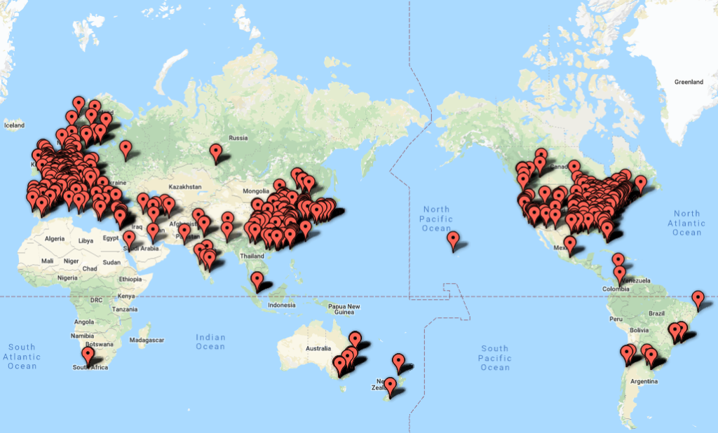 Fig. 5 A distributed map of BrainNet Viewer users
Download
The BrainNet Viewer is freely available on the NITRC website: www.nitrc.org/projects/bnv. If any questions, please visit https://www.nitrc.org/forum/forum.php?forum_id=2178 for help.
Citation
The following article should be referenced while using this tool.
Xia M, Wang J, He Y (2013) BrainNet Viewer: a network visualization tool for human brain connectomics. PLoS ONE 8(7): e68910. [ESI Highly Cited Paper] [Citations: 3100+] [PLOS ONE 10 Year Anniversary: 10 Highly Cited Articles] [PDF]